Anti-RSL1D1
Rabbit Polyclonal Antibody
Catalog No. R76-363R
| Catalog No. | Pack Size | Price (USD) | |
|---|---|---|---|
| R76-363R-100 | 100 ug | $675 | |
| R76-363R-BULK | BULK | Contact Us |
Rabbit Polyclonal Antibody
Catalog No. R76-363R
| Catalog No. | Pack Size | Price (USD) | |
|---|---|---|---|
| R76-363R-100 | 100 ug | $675 | |
| R76-363R-BULK | BULK | Contact Us |
Overview:
This antibody is designed, produced, and validated as part of a collaboration between Rockland and the National Cancer Institute (NCI) and is suitable for Cancer, Immunology and Nuclear Signaling research. PBK1 protein (also known as Ribosomal L1 domain-containing protein 1, cellular senescence-inhibited gene protein, and CATX-11) was isolated from highly invasive first trimester trophoblast cells and has been proposed to regulate their naturally occurring invasive behavior (Huch et al., 1998). PBK1 was also found to be over-expressed in non-small-cell lung cancer (NSCLC) cells (Petroziello et al., 2004). A recent study suggests that PBK1 may up-regulate the urokinase-type plasminogen activator (uPA) gene, which plays an important role in cellular matrix degradation and activation of other protease systems involved in cell invasion (Tong et al., 2005).
References:
1. Huch, G., et al.: Identification of differentially expressed genes in human trophoblast cells by differential-display RT-PCR. Placenta. 1998: 19 (8); 557-567.
2. Petroziello, et al.: Suppression subtractive hybridization and expression profiling identifies a unique set of genes overexpressed in non-small-cell lung cancer. Oncogene. 2004: 23 (46); 7734-7745.
3. Tong, C., et al.: Identification of a novel nucleus protein involved in the regulation of urokinase in 95D cells. Acta Biochim. Biophys. Sin (Shanghai). 2005: 37 (5); 303-309.
Specificity:
Recognizes the human PBK1 protein
Cross Reactivity:
Human
Host Isotype / Clone#:
Rabbit, IgG
Immunogen:
The antibody was produced against synthesized peptide corresponding to an internal sequence of human PBK1.
Purification:
Immunoaffinity chromatography
Stability:
1yr at –20oC from date of shipment.
Sample Data:
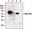
Western blot using Anti-RSL1D1 antibody shows detection of over-expressed PBK1 in lysates from HeLa cells transfected with Flag-PBK1. Lanes 1 and 3 contain lysate from Flag-PBK1 transfected HeLa cells. Lanes 2 and 4 contain lysate from cells transfected with null vector. Lanes 1 and 2 were blotted with anti-Flag antibody. Lanes 3 and 4 were probed with a 1:500 dilution of anti-PBK1. The band at 75 kDa, indicated by the arrowhead, corresponds to PBK1.
|
There are no related publications available for this product.
STAY CONNECTED
Fax: 1-604-232-4601


 Toll Free: 1-866-954-6273
 info@signalchem.com